Flat feet in children and adolescents: when to observe, when to treat, and what the evidence shows
In most children a flat foot is a shape, not a disease: the arch matures on its own in the first decade. What decides is whether the foot is flexible and whether it hurts. What the insole trials showed, how a rigid foot differs from a flexible one, when surgery is discussed at all, and what to ask the orthopaedic surgeon.
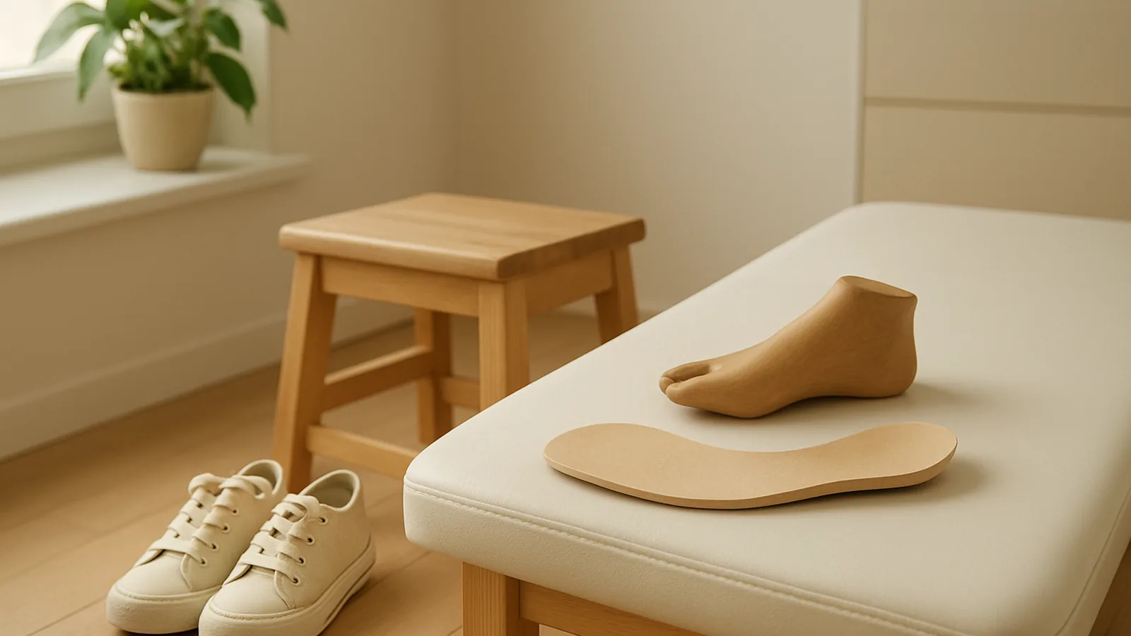
A flat foot in a child is one of the most common reasons for a visit to an orthopaedic surgeon — and one of the most common reasons to buy insoles. Yet for most children a flat foot is not a disease but a shape that changes on its own with growth. The question worth asking is not "is the foot flat" but "is it flexible, and does it hurt". Below is what the studies showed: who took part, what was measured, and what these papers do not say.
The arch matures: numbers by age
In a survey of 882 feet in 441 healthy people aged one to eighty, flat feet were usual in infants, common in children and fell within the range seen in normal adult feet; the authors recommended documentation and observation for a flexible flat foot that falls within that range (cross-sectional study, PMID 3818704).
In an Austrian study of 835 children aged 3–6, feet were scanned with a laser while standing. A flexible flat foot was found in 44 % of the children, and the proportion fell with age: 54 % at three years and 24 % at six. Boys had it more often than girls (52 % versus 36 %), and overweight children more often than normal-weight children. A pathological flat foot was found in less than 1 %, while 10 % of the children were wearing arch supports; by the authors' estimate, more than 90 % of the treatments given in this group were unnecessary (PMID 16882817).
In adolescents the numbers are different. In a medical database of 825,964 seventeen-year- olds in Israel, mild flexible flat foot was recorded in 12.4 % of males and 9.3 % of females, and severe flat foot in 3.8 % and 2.4 % respectively. Overweight and obesity were associated with a higher likelihood of flat foot (for obesity, odds ratio 1.77 in males and 1.55 in females), and greater height with a lower one (PMID 23696185). These are cross-sectional data from a conscription database: they show in whom flat feet are more common, but not what is cause and what is effect.
Hence the first conclusion: in a young child a flat foot is expected, and much of the arch forms during the first decade of life without any intervention. In a teenager the foot will no longer "mature" — but even there, the shape alone, without symptoms, is not a reason for treatment.
Flexible or rigid — the main dividing line
The clinician separates two fundamentally different conditions, and separates them with the hands, not with an X-ray. Flexible flat foot: the arch appears when the child rises onto tiptoe or sits with the foot unloaded, the heel turns inward on tiptoe, and the joints of the foot move freely. Rigid: the arch does not appear in any position, motion at the subtalar joint is limited and often painful.
A rigid flat foot in a teenager is a reason to look for a cause, and the most common one is tarsal coalition — a congenital fusion of two bones of the midfoot or hindfoot. Earlier work put its prevalence anywhere from 0.03 to 13 % depending on the method; in a series of 839 patients who had CT of both feet for various reasons at one Korean hospital, a coalition was found in 6.0 %, bilateral in 56.5 % of those, and the talocalcaneal type produced symptoms more often than the calcaneonavicular type (retrospective series, PMID 36513745). Reviews of paediatric orthopaedics describe a rigid or painful flat foot as a manifestation of coalition, collagen disorders, neurological disease and other conditions that need their own work-up — in contrast to the flexible form, which most authors consider a normal variant (PMID 24346183).
There is also a third variant that is easy to miss: a flexible flat foot with a short Achilles tendon. It was described as far back as 1948 (PMID 18921631), and it is this form, unlike the simple flexible one, that is known to cause pain in some adolescents and adults (review, PMID 21455468). The check is simple — how far the foot bends toward the shin with the knee straight.
What an X-ray measures and what it does not decide
A weight-bearing X-ray of the feet measures angles: the pitch of the heel bone, the angle between the talus and the first metatarsal, the height of the arch. These numbers are needed when the degree of deformity has to be documented or an operation planned; a coalition is sought differently — with an oblique view, indirect signs and, if needed, CT or MRI. But they do not make the decision "treat or observe": that decision is made on pain, function and flexibility. Tellingly, in the only randomised trial in which children were followed for three years by X-ray alone, the angles improved significantly in every group, including the untreated control group (PMID 2663868). Growth changes the film by itself.
Insoles and shoes: what the trials showed
This is the best-studied question, and the direction of the answer has not changed since 1989 — although the Cochrane review rated the certainty of the data themselves as low.
The Texas trial, 1989. 129 children with radiographically confirmed flexible flat foot, referred by paediatricians, were randomly assigned to four groups: observation, corrective orthopaedic shoes, a heel cup, or a custom-moulded plastic insert. Treatment lasted at least three years; 98 children with documented adherence completed the study. Radiographic angles improved in every group, including the controls, with no significant difference between treated and untreated children. The authors' conclusion: wearing corrective shoes or inserts for three years does not influence the course of flexible flat foot in children (RCT, PMID 2663868).
The Australian trial, 2007. 178 children aged 7–11 with bilateral flexible excess pronation were randomised to custom-made orthoses, ready-made orthoses or control; 160 finished. Gross motor proficiency, self-perception, exercise efficiency and pain were measured at baseline, 3 and 12 months. No group comparison was statistically significant on any outcome, including in the subgroup of children who presented with pain (RCT, PMID 17592702).
The Cochrane review, 2022. It included 16 randomised trials with 1,058 children aged 11 months to 19 years with flexible flat feet; the interventions were orthoses, footwear, exercises and electrical stimulation. The data were not pooled because of heterogeneity; no trial blinded its participants. In asymptomatic children, orthoses — custom or prefabricated — compared with ordinary shoes made little or no difference to the proportion free of pain at one year (one trial per comparison, about 106 participants each); function and quality of life were not measured in those trials. In children with juvenile idiopathic arthritis, custom orthoses in a small trial (28 participants, three months) were associated with better function; the authors rated the certainty of this evidence as low. The review's closing statement is blunt: in the absence of pain, the use of high-cost custom orthoses for healthy children with flexible flat feet has no supporting evidence, and the authors proposed retiring the research agenda on asymptomatic flat feet in healthy children altogether and redirecting it to conditions that actually cause pain and limit function (PMID 35080267).
What matters here is to read not only "does not help" but "in whom". The trials were done on flexible flat feet in otherwise healthy children. About rigid feet, coalitions or neurological causes these studies say nothing — there the approach is different and is decided separately.
On footwear there are also observational data. Among 2,300 children aged 4–13 in India, flat foot on static footprints was found in 8.6 % of those who wore shoes and 2.8 % of those who went barefoot; it was most common in children wearing closed-toe shoes, less common with sandals or slippers, and least common in the unshod (PMID 1624509). This is an association, not proof of an effect: the study is cross-sectional, and the groups may have differed in other ways. But it illustrates the overall conclusion of the reviews well: there is no evidence that an arch can be created in a child's foot by any external force or device (PMID 21455468).
Weight: the association exists, the effect of losing it has not been measured
The association between excess weight and flat foot reproduces across ages and countries: in preschoolers in Austria (PMID 16882817), in seventeen-year-olds in Israel (PMID 23696185), and in a systematic review of observational studies in children under ten, where obesity was associated with flat-footedness across the four included studies (PMID 32808447).
What these data do not contain is an answer to whether losing weight changes the shape of the foot, or the pain in it, in a child or adolescent. We did not find randomised trials with that outcome, and longitudinal data are limited to a few small series. So the phrase "it will go away once the weight comes off" remains a supposition, not a result — and that is a separate reason not to postpone the conversation about symptoms until "later".
Exercise and stretching
There are many trials of exercise for flat feet, but almost all were done in adults, last weeks, and measure intermediate quantities — plantar pressure distribution, navicular height — rather than pain and function in children. The Cochrane review included exercise trials in children but did not pool them and drew no separate conclusion about them (PMID 35080267).
The one situation where stretching is named directly in the professional literature is a flexible flat foot with an Achilles tendon contracture: a review from the American Academy of Orthopaedic Surgeons states that in these cases a stretching programme may help relieve pain, and in the same place notes that scant convincing evidence supports inserts or shoe modifications for relief of symptoms, and that there is no evidence those devices change the shape of the foot (PMID 25281257). That is an expert review, not a trial, and should be read as such.
What is known about the future: does a flat foot predict pain
What usually worries parents is not the shape of the foot now but what will happen in twenty years. The honest answer: we did not find longitudinal studies that followed an asymptomatic flexible flat foot from childhood into adult life with "pain" or "disability" as the outcome. A 2016 review in Pediatrics says so directly: there are no large prospective studies comparing the natural history of idiopathic flexible flat feet under different treatments, and the available literature does not identify which children are at risk of pain and disability as young adults; at the same time the authors call it safe and appropriate to simply observe an asymptomatic child with flat feet (PMID 26908688). Indirect data exist, and they disagree.
In 3,378 adult participants of the Framingham study, a planus foot posture was associated with arch pain in men (odds ratio 1.38), and pronated foot function with generalised foot pain (1.28) and heel pain (1.54) in men; in women no significant association between planus posture and more pain was found — on the contrary, a high arch and supinated function were associated with less pain. These are cross-sectional data adjusted for age and weight: they speak of association, not cause (PMID 23861176).
In military cohorts the picture is different. Among 246 US Army infantry trainees followed through 12 weeks of intensive training, the lowest rate of lower-limb injury was in the 20 % with the flattest feet, while the 20 % with the highest arches had an odds ratio for injury of 6.1 (PMID 7906597). In another cohort — 449 trainees at a Naval Special Warfare training centre — dynamic pes planus, pes cavus, restricted ankle dorsiflexion and increased hindfoot inversion were all identified as risk factors for overuse injury (PMID 10496574). Two studies in similar populations with different conclusions — that is the level of certainty available today.
Surgery: when it is discussed at all
The indications for surgery in flexible flat foot are stated strictly in paediatric orthopaedics: failure of prolonged non-surgical attempts to relieve pain that interferes with normal activities and is located under the medial midfoot or in the sinus tarsi; in nearly all such cases there is a contracture of the heel cord (review, PMID 25281257). An asymptomatic flat foot is not named as an indication in any of the reviews and guidelines we read.
What is known about the methods. The classic series of calcaneal lengthening — 31 feet in 20 children aged 4–16 with severe deformity resistant to conservative treatment, 26 of the 31 deformities being secondary to neuromuscular disorders — eliminated pain and callus in almost all feet at two to three and a half years of follow-up (case series, PMID 7713966). That is a result in the most severely affected patients, not in a teenager with a flexible foot and no pain.
Subtalar arthroereisis — placing an implant in the sinus tarsi — is widespread in Europe. A critical review of 76 publications up to 2010 showed improvement in most radiographic parameters, but complication rates of 4.8 to 18.6 % and unplanned implant removal rates of 7.1 to 19.3 %; the entire evidence base consisted of case series (PMID 22381197). A 2026 meta-analysis of 60 studies and 4,555 feet compared arthroereisis (4,089 feet) with osteotomies (448 feet) in children aged 2–18 with symptomatic idiopathic flexible flat foot: the radiographic angles changed differently, functional scores improved similarly (29.2 versus 26.4 points on the AOFAS scale), and complications were 9.2 % and 10.5 % respectively. There was one direct randomised comparison among the included studies — 66 patients, arthroereisis versus calcaneal lengthening; everything else was before-and-after series (PMID 41746382). In other words, the operations change angles and, according to case series, reduce pain in those who had it — but the comparison with conservative management still rests on observational data.
Transverse flat foot and hallux valgus in adolescents
A separate story is deformity of the forefoot: splaying of the metatarsals and outward deviation of the big toe. In a systematic review of nine studies (140 patients, 201 osteotomies, mean age at operation 14.5 years; among those operated on there were roughly ten girls for every boy — the make-up of the series, not a population frequency) the authors noted that non-operative management has a limited role in preventing progression, while contemporary operations give good satisfaction rates (86 %) with recurrence lower than historically reported; at the same time the whole literature is retrospective and heterogeneous (PMID 25899450). Here the decision is made on pain and on the pace of progression, not on an angle on the film.
What this changes in the conversation with the doctor
None of the above is a prescription: the approach to a flat foot is decided by a paediatric orthopaedic surgeon on examination. But the list of questions worth asking follows directly from these studies:
Is the foot flexible or rigid? Does the arch appear on tiptoe and when unloaded. This is the first question, and it matters more than any angle.
Does it hurt — where, after what kind of load, is there pain at rest or at night. An asymptomatic flexible foot is observed; a painful one is investigated.
Does the foot bend far enough toward the shin with the knee straight. A short Achilles tendon is a separate variant with a different approach.
If the foot is rigid, one-sided or painful in a teenager — has a coalition been excluded (oblique X-ray view, CT if needed).
If insoles are offered — for what exactly: for comfort with pain is one decision; "to correct the arch" is what did not happen in the trials.
If surgery is offered — what conservative treatment was tried and for how long, which technique exactly, and what is known about implant removal rates.
The red flags under which observation is not appropriate are named the same way across reviews: rigidity, one-sidedness, pain, progressive deformity, neurological signs, a marked Achilles contracture. Night pain is atypical for a flat foot altogether and calls for a search for another cause.
This is also where the usefulness of Lonevi ends: the record shows which measurements were taken and how they changed — including the films and reports brought back from orthopaedic visits. We do not prescribe treatment and we do not evaluate it; we help keep the series from which the trend is visible. There is a separate piece on how to show such a series to a doctor: sharing your record with a doctor. And on what the summary of a record does not do — limits of the summary.
This material is for information only and does not replace a consultation with a doctor. The diagnosis and management of a flat foot in a child are decided by a paediatric orthopaedic surgeon.
Articles in this section are educational and are not medical advice, a diagnosis, or a prescription. Consult a qualified professional before acting on anything you read here.
Read next
 ExplainerBiomarkers
What limits VO2max — lungs, heart or muscles — and how accurate the estimates are
VO2max is often described as a measure of the lungs. In healthy people the ceiling is set mostly by how much oxygen the heart and blood can deliver; the lungs become the limit only in particular situations. What changes with age, and how accurate lab tests, formulas and watches are.
12 min read ExplainerSleep
Mouth breathing during sleep in children: why it is not a habit, and what the treatment evidence shows
An open mouth at night usually means breathing through the nose or throat is harder, but the sign alone does not make a diagnosis. How sleep-disordered breathing in children and teenagers is confirmed, what trials of adenotonsillectomy, medicines, nasal treatment, orthodontics and exercises found, and where evidence is still missing.
15 min read ExplainerBiomarkers
Genetic age, telomeres and epigenetic clocks: what the tests actually measure
Three different things are sold as "genetic age": the inherited DNA sequence, telomere length and DNA methylation. What each one shows, how reproducible the measurement is, and what these tests do not say.
11 min read